
- Product introduction
- Detailed parameters
- Related resources
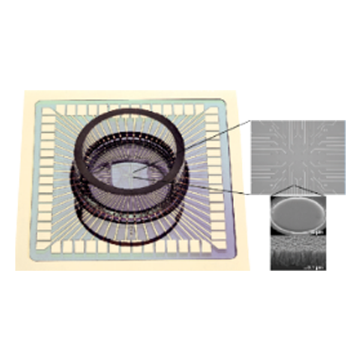
Located in germany Reutlingen institute of natural and medical sciences ( NMI)(www.nmi.de) is a research institute that produces high quality products using the most biocompatible materials MEA。NMI and Multi Channel Systems collaborated on several projects over the years.
Advanced quality control and production processes ensure MEA excellent consistent quality throughout. the electrode surface is coated with titanium nitride ( TiN), this is a very stable material that guarantees MEA can be reused many times.
Most MEA features a glass support which facilitates viewing of samples under any upright or inverted microscope. integrated circuits and contact pads are available in opaque and transparent versions, most MEA there is a built-in reference electrode.
Feature:
* Ics and contact pads available in opaque (titanium) or transparent (indium tin oxide)
* Long life and can be reused many times
* Electrode layouts for all application ranges
* Resistors have extremely low impedance
* Almost all MEA the reference electrode is integrated on the substrate of
* Electrode diameter as small as 8 microns
MEA- layout
Standard 8 x8 layout
8×8the electrode configuration is the most versatile. applications range from neuronal networks to brain slices, from stem cell-derived cardiomyocytes to heart cellstissue preparation. the electrode spacing can be100and200µmtwo types, provided separately700µmand1.4mmsquare recording area.
The diameter of the electrode is 10 µmand30µm,30µmadvantages of diameter electrodesyestheir low impedance and low noise levels,10µmelectrodes canrecord from single neurons or single cardiomyocytes.
ManyMEAfeatures a built-in reference electrode that allows cultures to be kept sterile during recording so thatlong-term recordings are performed on the culture and stimulation can also be performed from each electrode.

6x10 layout
6×10the distance between the electrodes of the layout is500µm,this creates4.5 mm × 2.5 mmrecording area, so thatrecord larger tissue samples on an array. each electrode can also be used for stimulation. all have6×10layoutMEAalso has an internal reference electrode. the electrode material isTiN。the micropillar structure of each electrode minimizes impedance and allows low-noise recording.extremely durable materials allow acute experiments up to50repeated cycles.
Hexagonal layout
* 60electrodes
* Available with same or different electrode diameters and distances
* Layout for retina recording
High density layout
* There are 60 in both recording areaselectrodes
* The electrode spacing is only30µm,the electrode diameter is only10µm
* High-resolution recording of individual neurons in neuronal networks
Special layout
* Various special electrode layouts developed together with customers
* Specially shaped stimulating electrodes or layouts with four high-density recording quadrants
* Layout can be customized according to needs
Porous layout* 60electrodes distributed to6in a hole
* 256electrodes distributed to9in a hole
* Exist24or96the hole has288electrode or in96the hole has1152porous plate with electrodes
* Applicable to toxicology, neurobiology, stem cell research and safety pharmacology
Applications:
MCSseveral are providedMEAgeometries and materials for various applications. almost any excitable or electrogenic cell and tissue can be used for extracellular recording in vitro, for example, central or peripheral neurons, cardiomyocytes, whole-heart specimen preparations, or the retina.
MEA- type
Perforation MEAs
Perforation MEA(pMEA)fabricated on thin polyimide foil, which is fixed to a glass carrier for physical stability. surrounding the electrode field is a circular area in which the foil is perforated (see image, black dots).


MEAdesigned to perfuse tissue on the array from the bottom. when usingMEAwhen the electrode records from an acute slice sample, the signal detected comes from cells at the bottom of the slice. these cells may be less healthy than cells on top because they receive less oxygen and nutrients from the perfusion solution. perfusion from the bottom solves this problem and may provide a better signal and improve long-term survival of acute slices. additionally, by usingMCSthe supplied constant vacuum pump applies negative pressure, allowing sections to beMEAthe surface remains in stable contact.
If you wish to usepMEA,all you need to do is create aMEA the system is equipped with a poured ground plate (PGP)and start recording.
With 256 electrodes MEAs
By reducing the electrode spacing, unique regions with higher spatial resolution can be mapped. exist16take16in the electrode array grid, we can obtain30,60,100and200µmelectrode spacing. for30µmspacing, electrode diameter is8µm,for60µmspacing, electrode diameter is10µm。 for100µmand200µmthe spacing uses30µmdiameter of the electrode. all256MEAlayouts all have four internal reference electrodes.
Features:
Higher spatial resolution
Larger recording area
Higher throughput
Thin MEAs
* Recording area with coverslip (180 µm) as thin
* Low working distance objectives for easy use of high magnification
* Uv transmission potential
* Transparent ic, perfect view
* Configurable with 60 or 256 electrodes
With 120 electrodes MEA
* Applicable to MEA2100- system
* 12X12 there are 120 electrodes in the grid of
* 4 internal reference electrode
* Suitable for stimulation
* Can be made into standard glass MEA and piercings MEA
EcoMEAs
* A low-cost option for routine experiments
* Gold electrodes (very strong and can be reused many times)
* 100 µm diameter electrode, 700 µm spacing
* Float glass carrier or printed circuit board
Pedot-CNT-MEAs
* Made of carbon nanotubes and PEDOT electrodes made of composite
* Repeatable low impedance
* High signal-to-noise ratio
* Good biocompatibility and cell adhesion
* Suitable for stimulation
Soft MEAs
* Made of soft polyimide foil (12 or 50 µm thick) made
* Available in 24, 36 and 72 electrodes
* For use in vivo /in vitro experiments and specific ex vivo uses
* Titanium nitride or gold electrode
* Can easily connect to MCS on the probe