- Product introduction
- Detailed parameters
- Related resources
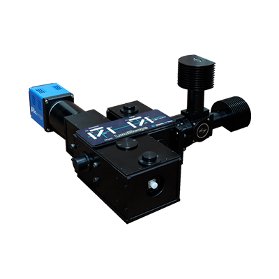
The system uses blue LED as the excitation illumination source, which is coupled to the deep brain region of the experimental animal through the GRIN lens (the cortex can be directly irradiated), and the induced green fluorescence is collected and transmitted by CMOS after bandpass filtering. The ultra-miniature microscope imaging system is suitable for commonly used laboratory model animals such as mice and rats.
Features:
*1mm diameter large field of view
*1mm working distance
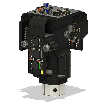
* Weight: 2.6g Height: 22mm
*+/-200um electronic autofocus
* Motion sensor to detect head steering activity
*Upgraded DAQ firmware and acquisition software
*Blue or green excitation light optional
The biggest advantage of the ultra-miniature microscopy system is that it can study the activity of brain nerve cells while the animal is moving freely. When the system is integrated with the behavioral analysis system, it is possible to collect both live nerve cell activity data and animal behavior data, which will help researchers better understand the function of the brain.