
- Product introduction
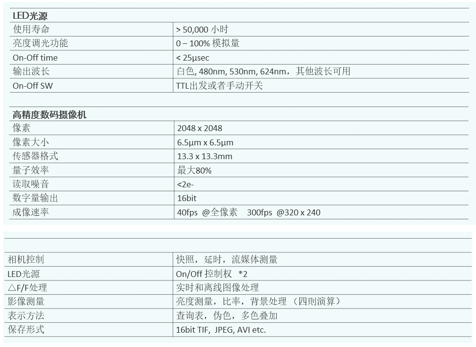
- Detailed parameters
- Related resources
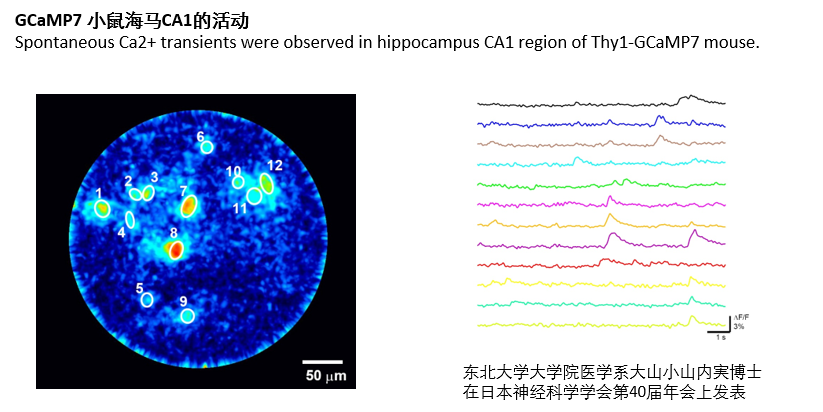
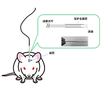
The LumiScope in vivo microscope uses an ultra-fine optical lens with a diameter of 350 μm and 10,000 imaging fibers arranged in an array to perform experiments with a variety of fluorescent proteins and fluorescent calcium indicators, as well as light-sensitive proteins such as ChR2 and NpHR, to achieve spatial resolution in the 2 μm range. The activity of specific nerves can be controlled by optogenetic stimulation.
The system is supported by an advanced model animal platform.
Features:
* Use GRIN lens and fiber optic for imaging
* minimally invasive deep brain imaging;
* Measurements under conditions of free movement of animals;
* Multiple brain regions were recorded at the same time;
* Compatible with a wide range of fluorescent proteins and fluorescent probes;
* Imaging of unlabeled neural activity by intrinsic fluorescence measurements;
* Capillary blood flow measurement;
* Fluorescence observation of deep organ cells in vivo;
* High-speed dual-wavelength fluorescence ratio imaging;
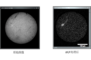
* The real-time/offline background processing of △F/F algorithm improves the signal-to-noise ratio;
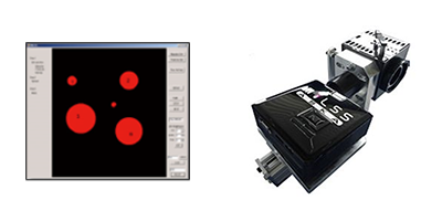
* Control and data analysis using multifunctional imaging software;
* Optogenetics irradiates multiple target brain regions at the same time.
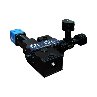
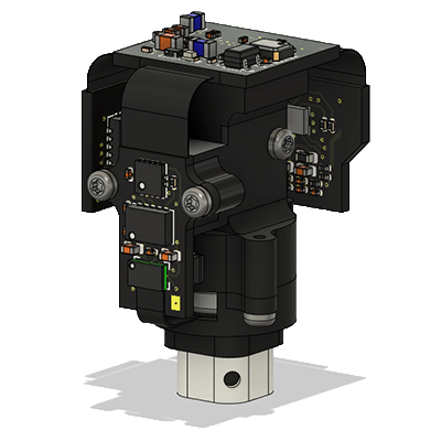
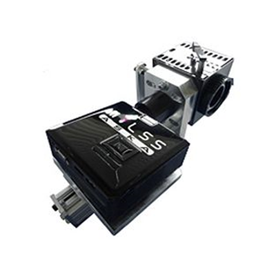
The LumiScope optical system part is an ultra-fine fluorescence endoscope (U-FEIS), which was developed by Lucille by Dr. Minoru Oyamauchi of the Graduate School of Medicine of Tohoku University in Japan with the assistance of JST and AMED Co., Ltd. is the result of the development.
Ultrafine fluorescence endoscope:
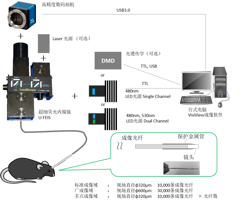
* LED, optogenetics input port x1, laser input port x1
* Camera mount C-mount
* Two fluorescence filter cubes can be mounted
* GFP filter set for SC or RFP filter set
* GFP/RFP Dual Bandpass Filter Kit for DC
* 2 sets of imaging fiber groups
* The diameter of the field of view is about Φ320μmL: 10,000 mm is about 10,000
* Array Imaging Fiber*1
* The outer diameter φ450-500μm is fixed on a metal protective tube*1
Optogenetics:
* The light source uses LEDs or lasers
* The irradiation time can be changed for each target
* Simultaneous illumination of several targets (up to 90 positions).
* Light of any wavelength is irradiated to each target
Multiple brain regions are imaged simultaneously
Multifield imaging fibers can be inserted into different sections to be measured, allowing photometry to be performed simultaneously using a single LumiScope. Up to 9 multipoint imaging fibers can be used.
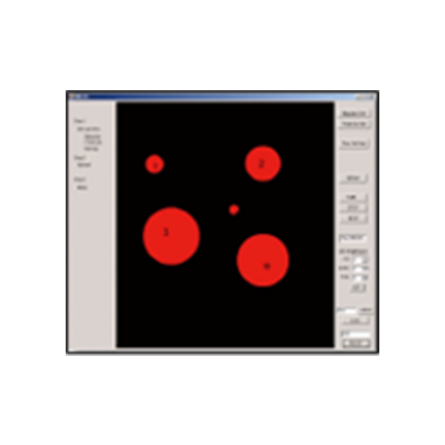
Imaging acquisition software
* Easy to set up and operate camera and LED light source control
* Multiple shooting modes snapshot, time-lapse, streaming, multi-color imaging
* Available online and offline. By ΔF/ Process the images obtained with improved signal-to-noise ratio
* Luminance analysis and ratio data processing are possible
Endoscopic fiber imaging microscope
* Cellular calcium imaging and optogenetics can be performed in multiple brain regions
* Multi-wavelength fluorescence imaging
* Records can be made on free-moving animals
Computer
* OS Windows10 64bit
* Desktop or laptop set
* 4ch TTL output
Imaging fibers and lenses are consumables and are not covered by the warranty
For laptops, an additional optional shutter controller is required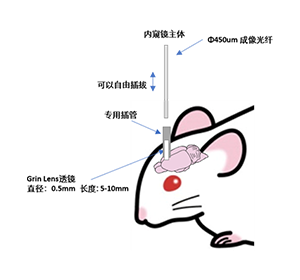
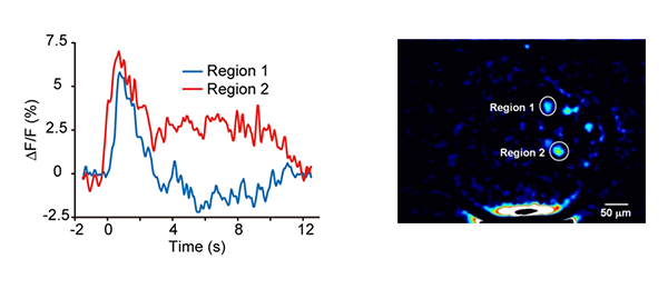
Application examples
Changes in neural activity associated with visual stimuli can be captured without labeling
Response when an endoscope is inserted into the cortical visual cortex and light stimulation is given to the other eye (fluorometric determination of flavin protein)